




Prototype Uncertainty-Driven Latent Diffusion for Semi-Supervised Medical Image Segmentation
-
摘要: 尽管半监督医学图像分割减少了对专家标注的依赖,但由于初始有标签样本稀缺,模型生成的伪标签常包含边界错误和拓扑畸变。这些错误在迭代训练中极易引发确认偏差,导致误差持续放大。为了解决该问题,提出一种基于原型不确定性驱动潜在扩散的半监督医学图像分割方法(Prototype Uncertainty-driven latent Diffusion, PU-Diff)。该方法包含两个优势:首先,利用高置信度预测样本构建类别原型,通过计算无标签像素与各类别原型的特征距离来量化分类不确定性,并生成不确定性引导图,从而精准定位伪标签中的潜在错误区域,为后续结构纠错提供指导;其次,将不确定性引导图与原始图像联合输入潜在扩散模型,在去噪过程中重点修复低置信度区域的边界与拓扑错误,从而增强模型对缺陷解剖结构的主动纠错能力,为参数优化提供更可靠的监督信号。实验表明,在ACDC、MS-CMRSeg、MSD Prostate和Kvasir-SEG四个数据集上,PU-Diff的分割性能均优于多种先进的半监督医学图像分割方法。Abstract:
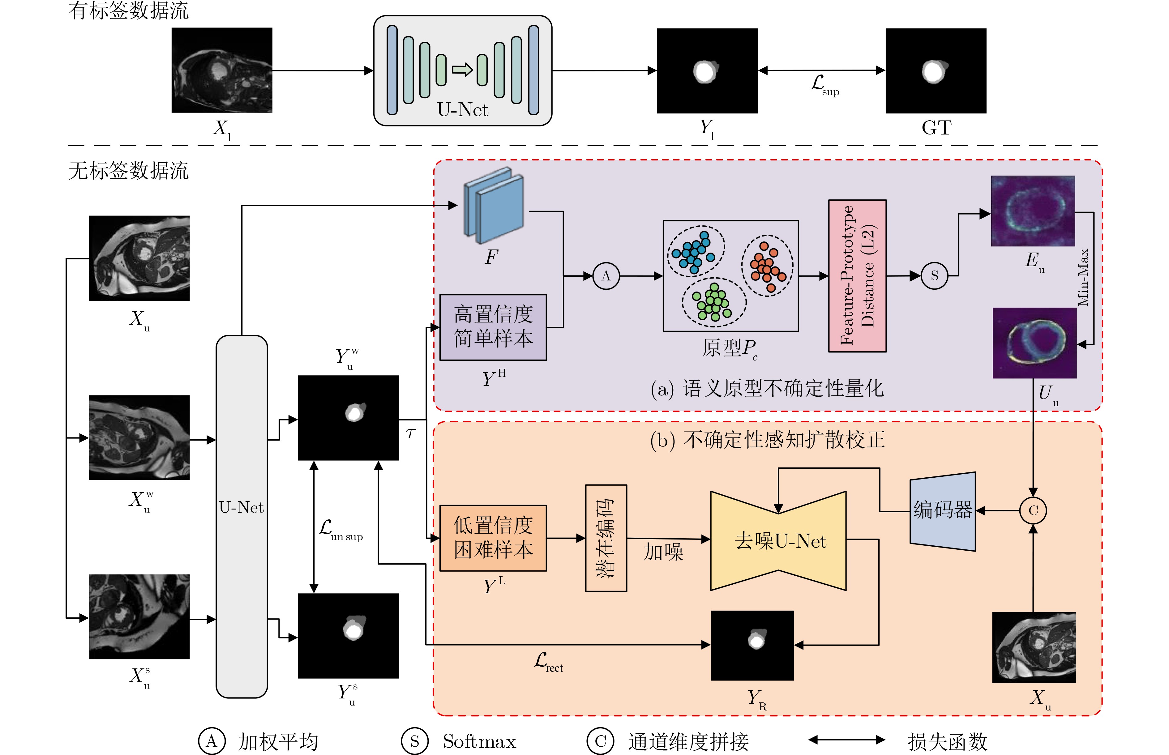
Objective Semi-supervised learning effectively alleviates the reliance on scarce pixel-level annotations in medical image segmentation, yet confirmation bias and error amplification caused by boundary errors and topological distortions in pseudo-labels severely limit the performance of conventional methods. Furthermore, traditional approaches often rely on shallow confidence scores, lacking the ability to explicitly reconstruct correct anatomical structures. This study develops a Prototype Uncertainty-driven latent Diffusion (PU-Diff) framework that utilizes semantic prototypes to quantify spatial uncertainty, explicitly forces the denoising process to rectify low-confidence regions, and aims to achieve precise error localization and active correction of complex anatomical structures under limited supervision. Methods Alongside the conventional consistency constraints, PU-Diff introduces an independent “predict, correct, and feedback” reverse path for pseudo-label rectification. Within this path, a Semantic Prototype Uncertainty Quantification (SPUQ) module constructs category prototypes utilizing high-confidence predictions. It then quantifies the classification ambiguity by calculating the distances in the high-dimensional feature space between unlabeled pixels and these prototypes, thereby generating a reliable spatial uncertainty guidance map. Subsequently, an Uncertainty-Aware Diffusion Rectification (UADR) module integrates this uncertainty map and the original image as joint spatial conditions to guide a latent diffusion model. This mechanism explicitly forces the denoising process to focus on repairing boundary and topological errors in low-confidence regions, ultimately generating high-fidelity pseudo-labels to provide reliable supervision signals for the main network's optimization. Results and Discussions Extensive experiments are conducted on ACDC, MS-CMRSeg, and MSD Prostate and Kvasir-SEG datasets. PU-Diff achieves clear performance gains. On the ACDC dataset with 5% and 10% labeled data, the average Dice coefficients reach 89.43% and 90.72%, respectively. On the MS-CMRSeg dataset with 10% labeled data, the average Dice reaches 88.06%, and on the MSD Prostate dataset with 10% labeled data, the average Dice reaches 72.64%, and on the Kvasir-SEG dataset with 10% labeled data, the average Dice reaches 86.61%. Ablation studies validate that the joint mechanism of SPUQ and UADR effectively eliminates structural noise and mends topological fractures. In terms of efficiency, PU-Diff achieves the optimal accuracy with a reasonable parameter count of 19.28 million and a single inference time of 3.56 ms, demonstrating a superior balance between segmentation accuracy and computational efficiency compared to existing advanced methods. Conclusions A semi-supervised medical image segmentation framework, PU-Diff, that integrates semantic prototype uncertainty quantification and latent diffusion-based label rectification is presented. PU-Diff mitigates confirmation bias by using high-dimensional feature prototypes to precisely locate error-prone areas, and addresses error accumulation by establishing an independent closed-loop that actively repairs topological distortions and boundary errors. Experiments on multiple medical imaging datasets show that PU-Diff achieves superior segmentation precision and structural fidelity compared with state-of-the-art semi-supervised methods, successfully maximizing the potential value of difficult unlabeled samples rather than passively discarding them. PU-Diff is architecturally adaptable and can be further optimized in computational complexity, facilitating its application to other complex anatomical structures, diverse imaging modalities, and clinical segmentation tasks under strict resource-constrained environments. -
表 1 不同数据集上PU-Diff(10%标注)与全监督U-Net的综合性能对比
数据集 U-Net[33] PU-Diff(10%标注) DSC (%) IoU (%) HD95 (mm) ASD (mm) DSC (%) IoU (%) HD95 (mm) ASD (mm) ACDC(均值) 91.42±0.36 84.57±0.54 1.37±0.11 0.94±0.09 90.72±0.68 82.96±0.51 1.58±0.23 0.97±0.19 MS-CMRSeg 88.21±0.45 79.32±0.49 3.90±0.33 1.21±0.16 88.06±0.43 79.13±0.32 4.33±0.61 1.56±0.35 MSD Prostate 73.92±0.50 61.45±0.41 7.33±0.24 1.80±0.13 72.64±4.14 60.15±4.57 7.18±2.02 1.99±0.27 Kvasir-SEG 89.14±0.27 84.35±0.33 2.23±0.16 1.40±0.08 86.61±0.35 83.10±0.41 3.11±0.20 2.22±0.13  下载: 导出CSV
表 2 在5%标注率的ACDC数据集上的对比实验
方法 RV Myo LV Mean DSC (%) IoU (%) HD95 (mm) DSC (%) IoU (%) HD95 (mm) DSC (%) IoU (%) HD95 (mm) DSC (%) Baseline 74.30±2.01 59.37±1.46 10.82±1.93 71.17±1.30 61.29±1.23 14.66±1.04 81.72±1.56 72.60±0.64 12.31±0.52 75.74±1.97 PatchCL[32] 79.67±1.74 68.49±2.85 7.82±2.79 79.25±1.62 70.82±1.48 8.73±1.55 90.41±2.13 83.27±1.81 9.94±1.75 83.11±1.42 BCP[16] 85.37±1.97 75.17±1.18 5.15±1.75 83.46±1.87 71.92±1.66 8.69±2.95 91.37±1.94 84.83±1.31 9.73±2.61 86.73±1.58 DPLR[17] 67.28±1.58 54.46±1.49 6.72±1.36 75.10±1.91 61.68±1.70 3.23±0.47 86.30±1.78 77.21±1.90 5.91±1.49 76.22±1.42 AD-MT[21] 84.58±1.50 74.32±1.64 3.88±0.83 86.55±1.64 76.51±1.57 2.91±0.92 91.13±1.63 84.74±1.42 4.08±1.63 87.42±1.64 SDCL[18] 87.09±1.46 77.31±1.53 2.43±0.47 84.04±1.55 72.98±1.45 2.49±0.74 91.43±0.81 83.71±1.57 3.60±0.44 87.52±0.94 DiffRect[24] 84.18±2.32 74.76±1.21 5.18±1.53 83.52±1.97 74.14±1.42 3.16±0.89 89.76±1.14 82.64±1.55 3.90±1.12 85.82±1.45 CGS[19] 85.45±2.51 73.97±1.71 6.77±1.32 84.31±0.94 73.23±1.02 3.03±0.56 90.96±0.56 84.07±1.12 2.89±0.71 86.90±0.83 PU-Diff 88.14±0.62 78.62±0.91 2.66±0.81 87.39±0.80 77.92±0.78 1.57±0.34 92.78±0.51 87.16±0.90 2.49±0.57 89.43±0.56 注:加粗数值表示最优值,下划线表示次优值。
下载: 导出CSV
表 3 在10%标注率的ACDC数据集上的对比实验
方法 RV Myo LV Mean DSC (%) IoU (%) HD95 (mm) DSC (%) IoU (%) HD95 (mm) DSC (%) IoU (%) HD95 (mm) DSC (%) Baseline 82.41±0.74 71.34±0.69 5.62±0.44 78.92±0.51 68.38±1.03 5.46±0.35 87.11±0.60 76.09±0.84 6.14±0.39 82.81±1.32 PatchCL[32] 86.09±2.19 76.27±1.97 2.86±1.03 83.11±1.75 71.38±1.81 2.14±1.74 90.15±1.77 82.87±1.95 3.57±1.80 86.45±1.49 BCP[16] 87.68±1.44 79.02±1.65 3.25±1.12 86.73±1.97 76.55±1.65 4.65±2.09 92.29±1.92 86.28±1.37 4.01±1.07 88.90±1.66 DPLR[17] 87.31±1.18 79.30±1.53 2.75±0.90 76.22±1.42 75.49±1.48 2.61±0.63 91.79±1.54 85.43±2.04 4.06±1.90 85.11±1.47 AD-MT[21] 88.73±1.84 80.42±1.50 1.48±0.62 87.42±1.54 76.76±1.24 1.49±0.37 92.98±1.85 87.25±1.12 2.91±0.74 89.71±1.73 SDCL[18] 89.37±1.51 81.47±0.87 1.55±0.47 87.52±1.68 78.12±0.92 1.38±0.19 93.18±1.88 87.19±0.94 2.95±0.49 90.02±1.40 DiffRect[24] 88.20±1.35 79.21±1.10 2.07±0.66 87.28±1.12 78.06±0.95 1.73±0.50 92.07±1.43 86.95±1.03 3.11±0.97 89.18±1.16 CGS[19] 87.97±1.09 78.67±0.92 2.52±0.69 86.90±0.85 76.00±0.71 2.43±0.52 91.32±0.43 84.86±0.44 3.29±0.95 88.73±0.83 PU-Diff 89.52±0.92 81.68±0.65 1.12±0.24 89.43±0.77 79.63±0.39 1.42±0.21 93.22±0.52 87.57±0.56 2.20±0.30 90.72±0.68 注:加粗数值表示最优值,下划线表示次优值。
下载: 导出CSV
表 4 不同方法在MS-CMRSeg数据集5%和10%标注比例下的对比实验结果
方法 5%标注比例 10%标注比例 DSC (%) IoU (%) HD95 (mm) ASD (mm) DSC (%) IoU (%) HD95 (mm) ASD (mm) Baseline 70.50±0.68 63.56±1.15 18.21±1.57 10.11±0.86 75.07±1.10 67.25±0.99 14.34±0.82 7.29±0.74 PatchCL[32] 79.94±1.71 70.13±1.64 14.43±2.28 7.34±1.40 83.71±1.62 72.39±1.44 11.73±2.16 5.84±1.42 BCP[16] 80.06±1.52 71.29±1.39 14.35±2.09 6.90±0.98 84.12±1.45 74.28±1.36 13.85±2.39 4.90±0.81 DPLR[17] 82.18±0.69 70.21±1.50 9.67±1.83 4.76±0.44 84.06±0.64 73.21±1.58 8.62±2.30 2.03±0.74 AD-MT[21] 83.86±0.94 73.02±0.97 9.91±0.80 3.17±0.65 85.83±0.97 76.97±0.93 8.81±0.94 1.99±0.38 SDCL[18] 84.66±0.74 74.25±0.64 8.49±1.63 3.03±0.57 86.75±0.72 76.60±0.54 7.93±1.08 2.97±0.84 DiffRect[24] 83.37±0.56 74.03±0.69 7.11±0.98 3.74±0.26 85.37±0.86 76.14±0.69 6.71±0.95 2.13±0.66 CGS[19] 84.05±0.59 73.51±0.37 8.29±0.42 4.30±0.88 84.92±0.57 75.01±0.42 7.99±0.92 3.90±0.89 PU-Diff 85.16±0.35 76.20±0.43 5.84±0.56 2.96±0.19 88.06±0.43 79.13±0.32 4.33±0.61 1.56±0.35 注:加粗数值表示最优值,下划线表示次优值。
下载: 导出CSV
表 5 不同方法在MSD Prostate数据集5%和10%标注比例下的对比实验结果
方法 5%标注比例 10%标注比例 DSC (%) IoU (%) HD95 (mm) ASD (mm) DSC (%) IoU (%) HD95 (mm) ASD (mm) Baseline 41.27±1.41* 31.96±2.60* 19.88±1.92* 13.76±1.68* 47.20±3.02* 35.17±4.12* 17.04±1.90* 10.83±0.89* PatchCL[32] 41.34±7.20* 32.68±9.75* 19.06±4.99* 13.17±4.24* 48.07±9.17* 35.80±10.58* 15.13±5.70* 10.45±4.21* BCP[16] 60.25±6.48* 48.83±7.46* 18.23±5.01* 9.92±2.70* 64.86±7.46* 50.04±8.63* 16.82±5.14* 8.31±2.98* DPLR[17] 56.14±7.91* 42.36±6.54* 21.97±6.86* 7.13±1.65* 59.60±8.40* 44.38±9.27* 20.51±7.25* 5.02±1.87* AD-MT[21] 69.25±6.38* 54.64±8.06* 8.78±2.30* 3.41±0.52* 71.42±6.38 57.73±8.46 7.55±2.74* 2.25±0.64* SDCL[18] 68.13±4.50* 57.04±4.21* 9.35±2.02* 3.76±0.63* 72.22±4.61* 59.30±5.70* 8.14±2.19 2.58±0.56* DiffRect[24] 62.14±4.12* 48.52±3.68* 13.08±4.14* 4.54±0.97* 63.94±5.19* 50.65±4.96* 10.14±2.11* 3.41±0.44* CGS[19] 64.76±5.25* 50.43±5.10* 10.27±2.87* 3.91±0.29* 66.93±7.23* 53.27±6.42* 9.11±2.98* 2.69±0.41* PU-Diff 69.71±2.66 58.89±3.27 8.14±1.32 2.96±0.30 72.64±4.14 60.15±4.57 7.18±2.02 1.99±0.27 注:粗体表示最优值,下划线表示次优值;*表示基于双边Wilcoxon符号秩检验,PU-Diff与其他方法的差异具有统计学显著性(p<0.05)。
下载: 导出CSV
表 6 不同方法在Kvasir-SEG数据集5%和10%标注比例下的对比实验结果
方法 5%标注比例 10%标注比例 DSC (%) IoU (%) HD95 (mm) ASD (mm) DSC (%) IoU (%) HD95 (mm) ASD (mm) Baseline 70.93±1.35 61.27±0.86 18.30±1.52 14.18±0.61 75.13±0.70 66.58±0.46 13.82±1.10 9.14±0.87 PatchCL[32] 74.51±2.74 64.40±2.32 16.17±2.16 10.54±2.07 80.98±2.25 73.84±1.76 13.41±1.05 7.36±1.20 BCP[16] 77.83±2.52 71.96±1.99 9.62±2.05 6.14±1.78 82.79±0.82 78.43±0.68 6.95±1.22 5.14±1.75 DPLR[17] 76.90±1.93 65.08±1.73 14.31±1.90 7.82±1.26 77.30±2.28 67.92±2.01 12.09±1.94 6.73±1.14 AD-MT[21] 80.34±1.05 77.47±1.28 8.84±1.52 5.98±0.95 81.64±1.26 79.70±1.34 6.42±1.19 5.24±0.32 SDCL[18] 83.59±0.87 80.46±0.68 6.73±0.79 4.04±0.62 85.06±0.57 82.75±0.39 3.97±0.23 3.02±0.21 DiffRect[24] 82.71±1.12 79.56±0.49 6.01±1.04 5.25±0.38 83.89±1.35 81.34±0.93 4.91±0.44 3.85±0.34 CGS[19] 79.04±0.28 78.31±0.40 7.93±0.52 5.18±0.46 82.37±0.26 80.77±0.80 5.36±0.23 4.16±0.30 PU-Diff 84.19±0.30 81.48±0.56 5.24±0.29 3.10±0.18 86.61±0.35 83.10±0.41 3.11±0.20 2.22±0.13 注:加粗数值表示最优值,下划线表示次优值。
下载: 导出CSV
表 7 UADR与SPUQ模块的消融实验
Version RV Myo LV DSC (%) IoU (%) HD95 (mm) DSC (%) IoU (%) HD95 (mm) DSC (%) IoU (%) HD95 (mm) Baseline 82.41±0.74 71.34±0.69 5.62±0.44 78.92±0.51 68.38±1.03 5.46±0.35 87.11±0.60 76.09±0.84 6.14±0.39 Baseline+UADR 86.70±0.49 77.63±0.51 2.61±0.37 85.68±0.94 77.69±0.77 2.62±0.30 91.12±1.58 85.97±0.79 3.43±0.93 Baseline+UADR +SPUQ 89.52±0.92 81.68±0.65 1.12±0.24 89.43±0.77 79.63±0.39 1.42±0.21 93.22±0.52 87.57±0.56 2.20±0.30
下载: 导出CSV
表 8 在MS-CMRSeg数据集上条件注入机制的消融
条件输入策略 DSC (%) IoU (%) HD95 (mm) ASD (mm) w/o Cond 84.29±0.67 75.66±0.42 10.02±1.17 5.37±0.91 I-only 85.86±0.49 77.42±0.56 9.73±1.03 4.49±0.52 U-only 87.23±0.51 78.59±0.44 7.10±0.96 3.73±0.40 I+U 88.81±0.38 80.30±0.58 4.21±0.89 1.53±0.18
下载: 导出CSV
-
[1] IQBAL S, KHAN T M, NAQVI S S, et al. TBConvL-Net: A hybrid deep learning architecture for robust medical image segmentation[J]. Pattern Recognition, 2025, 158: 111028. doi: 10.1016/j.patcog.2024.111028. [2] 阮东升, 施哲彬, 王嘉辉, 等. 结合跨模态特征激励与双分支交叉注意力融合的左心房疤痕分割方法[J]. 电子与信息学报, 2025, 47(5): 1596–1608. doi: 10.11999/JEIT240775.RUAN Dongsheng, SHI Zhebin, WANG Jiahui, et al. Left atrial scar segmentation method combining cross-modal feature excitation and dual branch cross attention fusion[J]. Journal of Electronics & Information Technology, 2025, 47(5): 1596–1608. doi: 10.11999/JEIT240775. [3] RAHMAN M, MUNIR M, and MARCULESCU R. EMCAD: Efficient multi-scale convolutional attention decoding for medical image segmentation[C]. 2024 IEEE/CVF Conference on Computer Vision and Pattern Recognition, Seattle, USA, 2024: 11769–11779. doi: 10.1109/CVPR52733.2024.01118. [4] DU Yuxin, BAI Fan, HUANG Tiejun, et al. SegVol: Universal and interactive volumetric medical image segmentation[C]. Proceedings of the 38th International Conference on Neural Information Processing Systems, Vancouver, Canada, 2024: 3516. [5] AZAD R, AGHDAM E K, RAULAND A, et al. Medical image segmentation review: The success of U-net[J]. IEEE Transactions on Pattern Analysis and Machine Intelligence, 2024, 46(12): 10076–10095. doi: 10.1109/TPAMI.2024.3435571. [6] GUO Xiaoyu, LIN Xian, YANG Xin, et al. UCTNet: Uncertainty-guided CNN-Transformer hybrid networks for medical image segmentation[J]. Pattern Recognition, 2024, 152: 110491. doi: 10.1016/j.patcog.2024.110491. [7] LI Zihan, ZHENG Yuan, SHAN Dandan, et al. ScribFormer: Transformer makes CNN work better for scribble-based medical image segmentation[J]. IEEE Transactions on Medical Imaging, 2024, 43(6): 2254–2265. doi: 10.1109/TMI.2024.3363190. [8] KARIMIJAFARBIGLOO S, AZAD R, KAZEROUNI A, et al. MedScale-Former: Self-guided multiscale transformer for medical image segmentation[J]. Medical Image Analysis, 2025, 103: 103554. doi: 10.1016/j.media.2025.103554. [9] ZHANG Yizhe. Medical image segmentation with minimal labeling effort: How far can we push the limits?[C]. Proceedings of the 40th AAAI Conference on Artificial Intelligence, Singapore, Singapore, 2026: 28492–28500. doi: 10.1609/aaai.v40i34.40079. [10] 刘振丙, 李焕兰, 王报源, 等. 融合客户端动态筛选的联邦半监督影像分割[J]. 电子与信息学报, 2026, 48(3): 960–970. doi: 10.11999/JEIT250834.LIU Zhenbing, LI Huanlan, WANG Baoyuan, et al. Federated semi-supervised image segmentation with dynamic client selection[J]. Journal of Electronics & Information Technology, 2026, 48(3): 960–970. doi: 10.11999/JEIT250834. [11] DU Xiaogang, WANG Dong, LEI Tao, et al. HGCL: Semi-supervised polyp segmentation via hierarchical granularity contrastive learning[C]. 2025 IEEE International Conference on Multimedia and Expo, Nantes, France, 2025: 1–6. doi: 10.1109/ICME59968.2025.11209964. [12] 樊亚文, 王潮远, 王鑫, 等. 融合反事实推理与轻量化设计的半监督脑肿瘤分割网络[J]. 电子与信息学报, 2026, 48(5): 2008–2019. doi: 10.11999/JEIT251130.FAN Yawen, WANG Chaoyuan, WANG Xin, et al. A lightweight semi-supervised brain tumor segmentation network with counterfactual reasoning[J]. Journal of Electronics & Information Technology, 2026, 48(5): 2008–2019. doi: 10.11999/JEIT251130. [13] RIZVE M N, DUARTE K, RAWAT Y S, et al. In defense of pseudo-labeling: An uncertainty-aware pseudo-label selection framework for semi-supervised learning[C]. Proceedings of the 9th International Conference on Learning Representations, 2021: 7533–7553. (查阅网上资料, 未找到对应的出版地信息, 请确认). [14] SU Jiawei, LUO Zhiming, LIAN Sheng, et al. Mutual learning with reliable pseudo label for semi-supervised medical image segmentation[J]. Medical Image Analysis, 2024, 94: 103111. doi: 10.1016/j.media.2024.103111. [15] 杨子瑶, 雷涛, 杜晓刚, 等. 基于可疑像素相互修正的半监督医学图像分割[J]. 电子学报, 2025, 53(5): 1607–1621. doi: 10.12263/DZXB.20240616.YANG Ziyao, LEI Tao, DU Xiaogang, et al. Semi-supervised medical image segmentation based on suspicious pixel mutual correction[J]. Acta Electronica Sinica, 2025, 53(5): 1607–1621. doi: 10.12263/DZXB.20240616. [16] BAI Yunhao, CHEN Duowen, LI Qingli, et al. Bidirectional copy-paste for semi-supervised medical image segmentation[C]. 2023 IEEE/CVF Conference on Computer Vision and Pattern Recognition, Vancouver, Canada, 2023: 11514–11524. doi: 10.1109/CVPR52729.2023.01108. [17] LU Yayuan, ZHANG Zheyu, ZHANG Yueyi, et al. Semi-supervised medical image segmentation via dynamic pseudo-label refinement[C]. 2024 IEEE International Symposium on Biomedical Imaging, Athens, Greece, 2024: 1–5. doi: 10.1109/ISBI56570.2024.10635316. [18] SONG Bentao and WANG Qingfeng. SDCL: Students discrepancy-informed correction learning for semi-supervised medical image segmentation[C]. Proceedings of the 27th International Conference on Medical Image Computing and Computer Assisted Intervention, Marrakesh, Morocco, 2024: 567–577. doi: 10.1007/978-3-031-72111-3_53. [19] WANG You, LI Zekun, QI Lei, et al. Balancing multi-target semi-supervised medical image segmentation with collaborative generalist and specialists[J]. IEEE Transactions on Medical Imaging, 2025, 44(7): 3025–3037. doi: 10.1109/TMI.2025.3557537. [20] HU Ming, YIN Jianfu, MA Zhuangzhuang, et al. β-FFT: Nonlinear interpolation and differentiated training strategies for semi-supervised medical image segmentation[C]. 2025 IEEE/CVF Conference on Computer Vision and Pattern Recognition, Nashville, USA, 2025: 30839–30849. doi: 10.1109/CVPR52734.2025.02872. [21] ZHAO Zhen, WANG Zicheng, WANG Longyue, et al. Alternate diverse teaching for semi-supervised medical image segmentation[C]. Proceedings of the 18th European Conference on Computer Vision, Milan, Italy, 2025: 227–243. doi: 10.1007/978-3-031-72652-1_14. [22] PAN Qingtao, QIAO Wenhao, LOU Jingjiao, et al. DuSSS: Dual semantic similarity-supervised vision-language model for semi-supervised medical image segmentation[C]. Proceedings of the 39th AAAI Conference on Artificial Intelligence, Philadelphia, USA, 2025: 6299–6307. doi: 10.1609/aaai.v39i6.32674. [23] WU Junde, FU Rao, FANG Huihui, et al. MedSegDiff: Medical image segmentation with diffusion probabilistic model[J]. Proceedings of Machine Learning Research, 2023, 227: 1623–1639. (查阅网上资料, 请核对文献类型及格式). [24] LIU Xinyu, LI Wuyang, and YUAN Yixuan. DiffRect: Latent diffusion label rectification for semi-supervised medical image segmentation[C]. Proceedings of the 27th International Conference on Medical Image Computing and Computer Assisted Intervention, Marrakesh, Morocco, 2024: 56–66. doi: 10.1007/978-3-031-72390-2_6. [25] CHEN Yunzhu, LIU Yang, LU Manti, et al. Multi-consistency for semi-supervised medical image segmentation via diffusion models[J]. Pattern Recognition, 2025, 161: 111216. doi: 10.1016/j.patcog.2024.111216. [26] ZHANG Le, WU Fuping, BRONIK K, et al. DiffuSeg: Domain-driven diffusion for medical image segmentation[J]. IEEE Journal of Biomedical and Health Informatics, 2025, 29(5): 3619–3631. doi: 10.1109/JBHI.2025.3526806. [27] BERNARD O, LALANDE A, ZOTTI C, et al. Deep learning techniques for automatic MRI cardiac multi-structures segmentation and diagnosis: Is the problem solved?[J]. IEEE Transactions on Medical Imaging, 2018, 37(11): 2514–2525. doi: 10.1109/TMI.2018.2837502. [28] ZHUANG Xiahai. Multivariate mixture model for myocardial segmentation combining multi-source images[J]. IEEE Transactions on Pattern Analysis and Machine Intelligence, 2019, 41(12): 2933–2946. doi: 10.1109/TPAMI.2018.2869576. [29] WU Fuping and ZHUANG Xiahai. Minimizing estimated risks on unlabeled data: A new formulation for semi-supervised medical image segmentation[J]. IEEE Transactions on Pattern Analysis and Machine Intelligence, 2023, 45(5): 6021–6036. doi: 10.1109/TPAMI.2022.3215186. [30] ANTONELLI M, REINKE A, BAKAS S, et al. The medical segmentation decathlon[J]. Nature Communications, 2022, 13(1): 4128. doi: 10.1038/s41467-022-30695-9. [31] JHA D, SMEDSRUD P H, RIEGLER M A, et al. Kvasir-SEG: A segmented polyp dataset[C]. Proceedings of the 26th International Conference on MultiMedia Modeling, Daejeon, South Korea, 2020: 451–462. doi: 10.1007/978-3-030-37734-2_37. [32] BASAK H and YIN Zhaozheng. Pseudo-label guided contrastive learning for semi-supervised medical image segmentation[C]. 2023 IEEE/CVF Conference on Computer Vision and Pattern Recognition, Vancouver, Canada, 2023: 19786–19797. doi: 10.1109/CVPR52729.2023.01895. [33] RONNEBERGER O, FISCHER P, and BROX T. U-Net: Convolutional networks for biomedical image segmentation[C]. Proceedings of the 18th International Conference on Medical Image Computing and Computer-Assisted Intervention, Munich, Germany, 2015: 234–241. doi: 10.1007/978-3-319-24574-4_28. -

 下载:
下载:



图(5) / 表(9)
计量
- 文章访问数: 14
- HTML全文浏览量: 2
- PDF下载量: 0
- 被引次数: 0
